The molecular mechanisms of retinal lamination
|
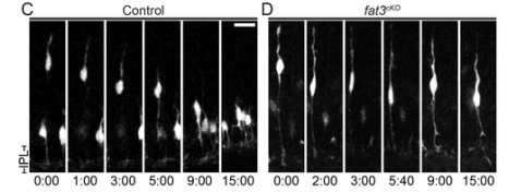
Most auditory information processing takes place in the brainstem; to convey this information rapidly, spiral ganglion neurons have a long, bipolar morphology with targets that are spaced far apart. In contrast to the auditory system, visual information undergoes abundant processing within the retina before reaching the retinal ganglion neurons. Hence, visual networks exhibit a fundamentally different type of organization, with cells and processes arranged in layers and making many local connections. To study how these layers form, we are analyzing mice with mutations in Fat3, an enormous atypical cadherin. In the absence of Fat3, amacrine cells extend an extra dendrite that give rise to an ectopic plexiform layer in the middle of the inner nuclear layer. In addition, an excess number of amacrine cells are displaced into the retinal ganglion cell layer. We are using a combination of biochemistry, retinal electroporations, live imaging, and mouse genetics to determine the cellular and molecular functions of Fat3 in retinal circuit assembly.
|


